
Fetal Ultrasound in Spartanburg, SC
Fetal ultrasound provides you and your doctor with a first glimpse of your developing baby. The highly trained OBGYNs at Spartanburg & Pelham OB-GYN have extensive experience performing fetal ultrasounds. Request an appointment at our office in Spartanburg or Greer, SC today!
Quick Facts
|
Types of Ultrasounds
-
 Standard: Directs high-pitched sounds waves toward the baby, which bounces off tissues, organs and bones in the
mother's body, including those of the baby in uterus. This creates black and white images on a
monitor.
Standard: Directs high-pitched sounds waves toward the baby, which bounces off tissues, organs and bones in the
mother's body, including those of the baby in uterus. This creates black and white images on a
monitor.
- Advanced or Targeted: Used to further investigate a suspected abnormality identified by a standard ultrasound.
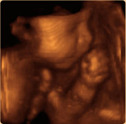
- Three-dimensional: Offers 3-D images with a high level of detail.
- Doppler: Measures slight changes in the frequency of the ultrasound waves as they bounce off moving objects, such as blood cells.
How To Prepare
- Wear comfortable, loose-fitting clothing.
- Most likely the test needs to be done with a full bladder.
- For transvaginal ultrasound or those in late pregnancy, a full bladder usually isn't necessary.
What To Expect
- The examination usually takes less than 30 minutes.
- The patient is usually positioned on an examination table and clear gel is applied to the abdomen. This improves conduction of sound waves and eliminates air between the transducer, a small plastic device that sends out sound waves and records them as they bounce back, and your skin.
- The transducer moves back and forth over the abdomen, directing sound waves into the uterus and capturing the reflected sounds waves that are digitally converted into images.